
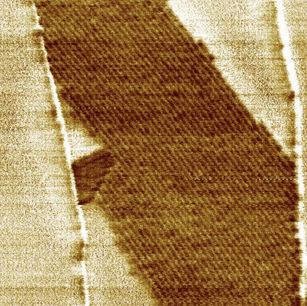
Application fields
Key Features
Phase Imaging
- Material Property Mapping: Quantify stiffness, adhesion, and viscoelastic differences with high sensitivity.
- High Spatial Resolution: Detect nanoscale variations in mechanical properties.
- Non-Destructive Imaging: Suitable for fragile and soft samples.
- Multimodal Integration: Combine with other AFM modes for comprehensive analysis.
- Versatility: Works in air, liquid, or controlled environments.
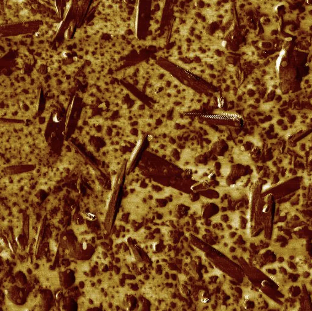
Phase imaging mode reveal surface structure and material property variations across a 2 µm area
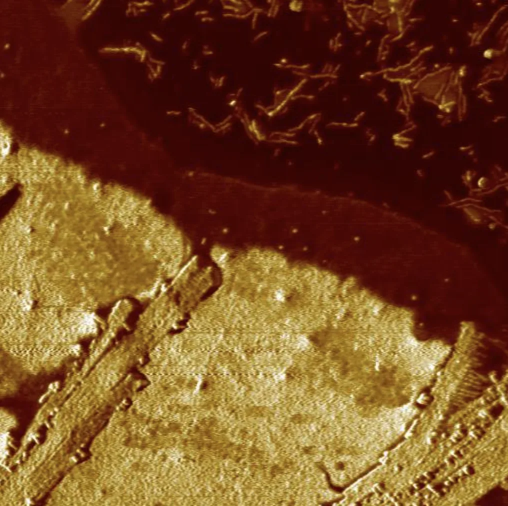
Applications of Phase Imaging
Polymers and Composites
- Map stiffness and adhesion variations in polymer blends.
- Analyze phase separation in composite materials.
Biological Samples
- Study the mechanical properties of cells and tissues.
- Investigate biomaterial surfaces for medical applications.
2D Materials and Nanotechnology
- Characterize nanoscale properties of graphene and other 2D materials.
- Evaluate the homogeneity of thin films and coatings.
How does Phase Imaging work?
Phase Imaging analyzes the phase shift between the probe oscillation and the drive signal as the tip interacts with the sample. This phase shift reflects differences in mechanical properties, such as adhesion and stiffness, enabling detailed mechanical property mapping at the nanoscale.
Specifications
- Resolution: Sub-nanometer in vertical resolution.
- Phase Sensitivity: Detects subtle variations in stiffness and adhesion.
- Operating Environments: Air, liquid, and controlled conditions.
- Applications: Polymers, nanocomposites, biological samples, and 2D materials.
Questions about Phase Imaging?
Ask the application scientists behind our AFMs and modes — about your samples, probes, set-up or which mode fits your measurement. We answer every message personally.